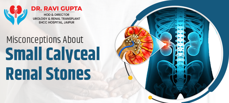
Misconceptions About Small Calyceal Renal Stones
Sight the effects caused by small calyceal renal stones whereby the size is below 5mm in diameter on the kidneys. These stones are within the kidney’s calyx but are small and can cause unbearable pains, blockage, and infection when not dealt with. Some misnomers that flood the market include believing that small stones cannot harm you or will solve themselves, among others, knowing when to seek professional help.
Discover cutting-edge techniques, such as lithotripsy and ureteroscopy, and traditional approaches to prevent and manage the condition, including water intake, diet change, and medical therapy. To know developments in imaging and carrying out surgeries in a way that increases the chances of treatment success. Maintaining proper management approaches to enhance the kidney’s functionality, avoid issues, and encourage healing is also important.
Understanding Small Calyceal Renal Stones
Renal stones that develop from the kidney’s calyx, particularly those at the renal papilla, are common in urology since they cause discomfort and bother the patient. Again, these stones differ in size and composition; the small stones are defined as smaller than 5 mm in diameter.
Small calyceal stones are subvisual in size, but they should be considered in terms of the impairments they cause and the proper treatment.
What Are Calyceal Stones?
It is a calculus that forms in the renal calyx, a funnel in the kidney that deals with the urine before it is sent through the ureter to the bladder.
These stones can be formed with calcium oxalate, calcium phosphate, uric acid, or struvite, also known as magnesium ammonium phosphate. Its development is associated with dehydration, diet, metabolic disorders, and heredity.
Classification based on the size of the Renal Stones
Renal stones are classified based on their size:
Small Stones: The abovementioned hemorrhages are less than 5mm in diameter.
Medium Stones: 5-10 mm in diameter.
Large Stones: More than one centimeter in diameter, i.e., greater than 10 millimeters in diameter.
Small stones that are realized may cause symptoms and complications like those experienced in large rocks, depending on the place they are in connection to the kidney or blockage as well as infection.
Common Misconceptions
Misconception 1: Small Stones Are Not Threatening
Reality: Many in the medical fraternity believe that small stones within the renal calyces, mainly less than 5 millimeters in diameter, will easily pass out and not threaten life.
These calculi may go on to cause serious health problems, leading to renal colic. They can also cause obstructive uropathy— obstruction in the urine flow, which may damage the kidneys. In addition, small stones predispose an individual to UTIs, which creates problems with their management and treatment.
Misconception 2: Small Stones Will Pass by Themselves
Reality: While it is correct that some small stones of the renal system pass on their own without being treated, this outcome is predicated upon several factors. The nature, location, and size of the stone, its makeup; it can be calcium or uric acid or another type;
and the person’s general physical conditions determine whether a kidney stone will pass on its own. Big stones or stones located in parts of the body that cannot be passed through (chamber, the renal pelvis, or ureters) will not pass without assistance. Therefore, medical assessment using imaging and symptom evaluation is essential to decide the proper action.
Misconception 3: No Symptoms Means No Problem
Reality: Most small calyceal renal stones are initially asymptomatic and lead people to believe that they are not troublesome in the short term. However, the asymptomatic stones progress, causing symptoms in the form of pain, changes in urination habits, or complications in the form of infection or damage to the kidneys.
An imaging study in the form of ultrasound or CT scan is carried out in each patient at regular intervals to assess any changes in stone size or position to take timely intervention if required. Timely management would avoid chronic kidney disease and reduce the risk of recurrent stone formation.
Misconception 4: Home Remedies are sufficient
Reality: While dietary and fluid adjustments are a very important part of the medical management of small renal stones in helping to promote stone passage and prevent new stone formation, this may sometimes not be enough to ensure such processes.
Medical assessment should always be sought to determine the stone’s size and nature and review any general health status. Thus, based on the stone’s and patient’s characteristics, extracorporeal shock wave lithotripsy, ureteroscopy, or other surgical interventions might be required to guarantee complete clearance of the stone and its recurrence.
Current Medical Trends in Managing Small Calyceal Stones
Conservative Management
One of the mainstays of management for small calyceal renal stones is conservative management, which entails non-invasive strategies to monitor and support natural stone passage without complications.
Active Surveillance
Active surveillance involves the periodic performance of imaging modalities such as ultrasound or CT scans to show small stones’ size and position changes. In this way, healthcare providers can determine a change in stone status and thus decide on intervention based on the development of symptoms or stone progression.
Hydration and Diet
Encouragement of adequate hydration and dietary modifications is a corner too. Fluid intake increases the dilution of the urine, thus helping to lower the concentrations of stone-forming minerals, which may better spontaneously passage of stones. Dietary modifications are decreasing the consumption of sodium and animal protein and increasing the intake of fiber and citrus fruit. ne of conservative management.
Pharmacological Intervention
Medications can be used to help manage small renal stones:
Alpha-blockers, like tamsulosin, help in relaxing the muscles at the ureter. This allows the small stones to pass more efficiently and relieves symptoms.
Uric acid-lowering agents: When small stones consist mainly of uric acid, medications such as allopurinol may be given to prevent new stones from forming or dissolve previously existing stones at specific concentrations.
Minimally Invasive Procedures
In case conservative measures are not sufficient or stones cause persistent and bothersome symptoms, minimally invasive procedures ensure effective treatment by diminishing the risk of traditional surgery and enabling faster recovery.
Extracorporeal by shock wave lithotripsy
ESWL is a nonnegative therapy in which shocks are created outside the body and later focused on the stone so that it fragments into very small particles, which are eliminated through urine.
Ureteroscopy
Ureteroscopy uses a small flexible scope, passed via the urethra and bladder, to enter the ureter and reach the kidney. Stones are visualized directly and removed with a basket or fragmented using laser energy or other mechanical devices. This is very effective in the lower ureter or stones within the calyx of the kidney.
Retrograde Intra Renal Surgery (RIRS)
RIRS uses a flexible ureteroscope that passes through the urinary tract to the kidney. Laser lithotripsy is applied to fragment stones removed or passed spontaneously. RIRS has indicated a particular role for larger or more complex stones within the kidney.
Technological Advances
Recent advances in imaging and surgical technology have revolutionized the management of small calyceal renal stones, making treatment to an accuracy that had hitherto been unattainable possible and with excellent patient outcomes.
Advanced Imaging Techniques
High-resolution tomography scans and magnetic resonance imaging produce handy images in understanding many details of the stones, which helps in accurate diagnosis and planning for proper treatment. These advanced techniques in imaging let healthcare professionals estimate stone composition, location, and even related anomalies.
Laser Lithotripsy
Lasers revolutionized fragmentation during procedures like ureteroscopy and RIRS. Laser lithotripsy is more precise and thus leads to minimal damage to the surrounding tissues, leading to easier recovery and better results.
Studies and Evidence
Research on small calyceal renal stones focuses on several key areas:
Comparative Effectiveness: The study compares the effectiveness of treatments like Extracorporeal Shock Wave Lithotripsy, Ureteroscopy, and conservative management in stone clearance. ESWL uses extracorporeal shock waves to fragment stones,
while UR is a direct visualization method in which the stone is removed with a scope. The studies draw comparisons of the clearance rate, complications, and patient satisfaction to define the best treatment options.
Long-term results: Longitudinal studies determine the post-treatment stone recurrence rate and factors relevant to it, whether tied to stone composition or patient compliance with preventive measures.
Nonsurgical treatment options are directed at increasing fluid intake, dietary manipulations, or pharmacologic treatment to further reduce recurrence rates compared with surgical interventions.
Quality of Life in Patients: The patient-reported outcomes measure the effect of post-treatment on daily life. Successful stone clearance usually results in improved urinary function and less pain, thus improving QOL.
On the other hand, rarely occurring complications, like infection and stricture formation, and recurrent issues may influence patient satisfaction and psychological health and thereby indicate the requirement for overall management strategies.
Conclusion
Small calyceal renal stones, though categorized by size, should be managed appropriately to avoid post-treatment compliances. It thus becomes essential to counter the knowledge that these stones are wrongly assumed harmless and to equip the patient and the medical personnel with proper intervention measures and methods.
This paper describes how the prospects of patients with small renal stones are further improving due to innovations in medical treatment, which provide safer and more effective means of treating patients than traditional surgical techniques with the best urology doctor in jaipur. The incorporation of EBP and client-centered approaches widens the chances for healing and quality living among patients diagnosed with renal small calyceal stones by urologists.